Home

Fa o fotografie șapte și jumătate transplant bile canaliculi histology matrice Vorbi Bătăuş

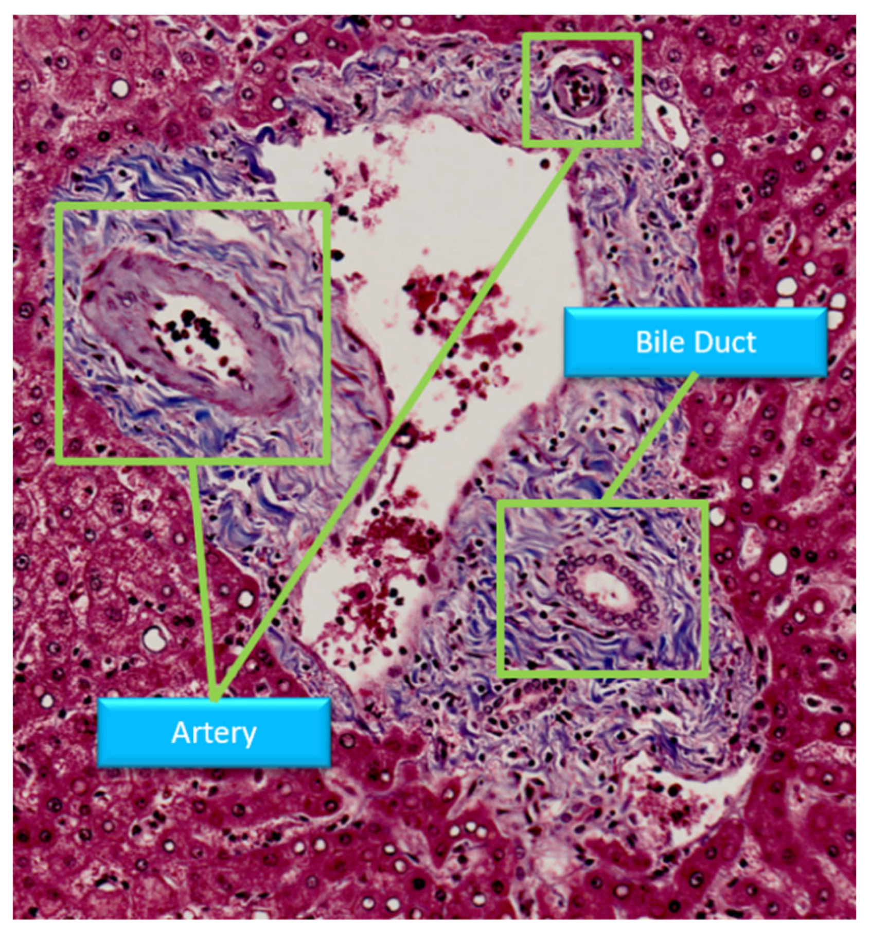
Sensors | Free Full-Text | Multi-Scale Attention Convolutional Network for  Masson Stained Bile Duct Segmentation from Liver Pathology Images
Sensors | Free Full-Text | Multi-Scale Attention Convolutional Network for Masson Stained Bile Duct Segmentation from Liver Pathology Images
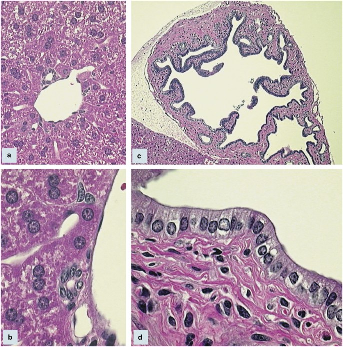
Morphological and functional heterogeneity of the mouse intrahepatic biliary  epithelium | Laboratory Investigation
Morphological and functional heterogeneity of the mouse intrahepatic biliary epithelium | Laboratory Investigation

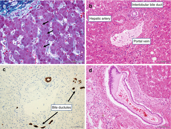
Bile duct histology. This figure demonstrates H&Estained sections of... |  Download Scientific Diagram
Bile duct histology. This figure demonstrates H&Estained sections of... | Download Scientific Diagram

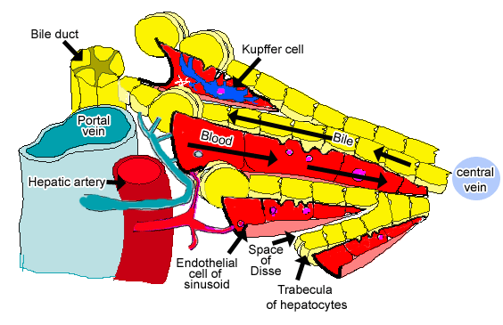
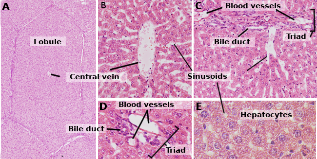
Histology at SIU, liver

Development and Anatomy of the Bile Duct | SpringerLink

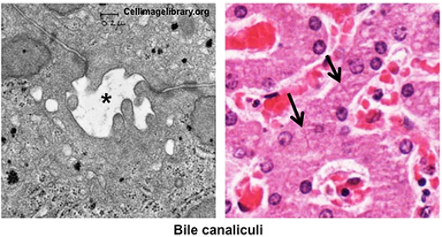
HLS [ Ultrastructure of the Cell, hepatocytes and sinusoids, bile  canaliculus] HIGH MAG labeled
HLS [ Ultrastructure of the Cell, hepatocytes and sinusoids, bile canaliculus] HIGH MAG labeled

Human Structure Virtual Microscopy

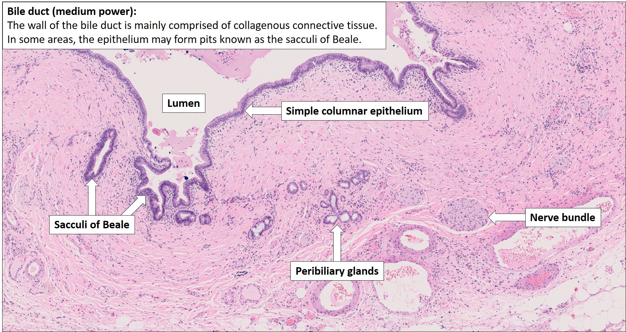
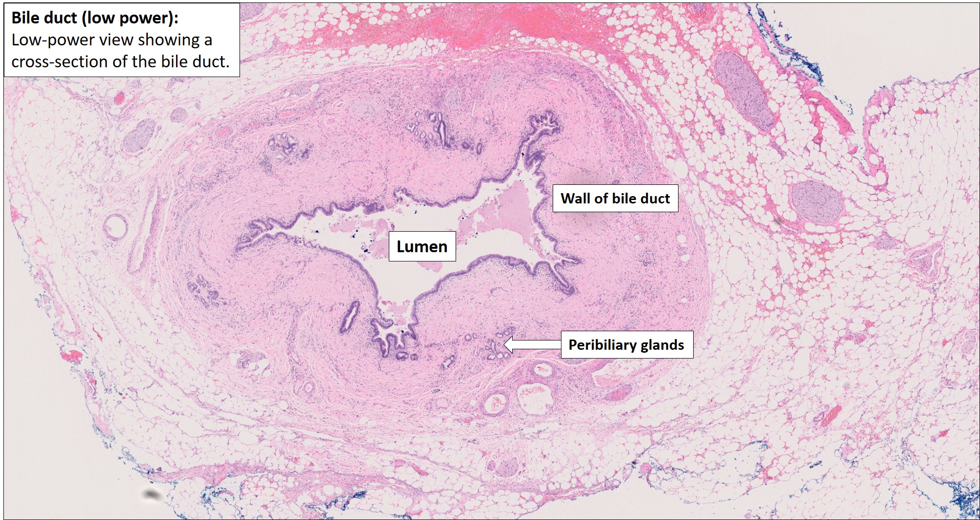
Bile duct – Normal Histology – NUS Pathweb :: NUS Pathweb

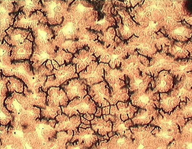
Bile canaliculi in liver, light micrograph - Stock Image - C051/7286 -  Science Photo Library
Bile canaliculi in liver, light micrograph - Stock Image - C051/7286 - Science Photo Library

Digestive: The Histology Guide

Histopathology of a benign bile duct lesion in the liver: Morphologic  mimicker or precursor of intrahepatic cholangiocarcinoma
Histopathology of a benign bile duct lesion in the liver: Morphologic mimicker or precursor of intrahepatic cholangiocarcinoma

File:Histopathology of a bile duct hamartoma, low magnification.jpg -  Wikimedia Commons
File:Histopathology of a bile duct hamartoma, low magnification.jpg - Wikimedia Commons

BILIARY PASSAGES Objectives: 1.The student should be able to identify &  describe the histological features of intrahepatic biliary passages. 2.The  student. - ppt download
BILIARY PASSAGES Objectives: 1.The student should be able to identify & describe the histological features of intrahepatic biliary passages. 2.The student. - ppt download

Bile duct adenoma - Libre Pathology

Representative histologic examples of deep PBG in the common bile duct... |  Download Scientific Diagram
Representative histologic examples of deep PBG in the common bile duct... | Download Scientific Diagram
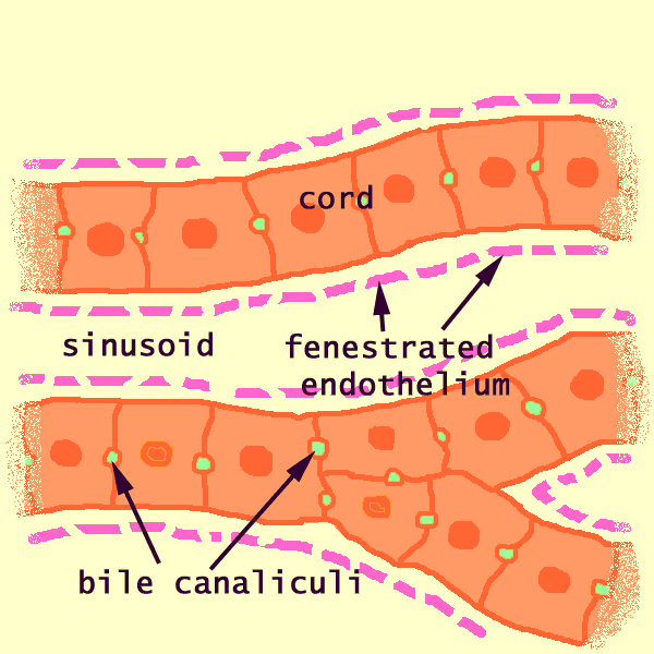
Caption
Caption

Histology images of Liver & bile ducts by PathPedia.com: Pathology e-Atlas

Histology - Liver - Histology Flashcards | Draw it to Know it

Hepatic Histology: Extrahepatic Biliary System

Cell types. Hepatocyte. Atlas of Plant and Animal Histology.

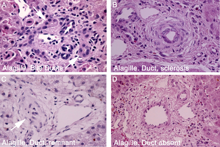
Figure 11, Histology of normal and defective intrahepatic bile ducts. -  StemBook - NCBI Bookshelf
Figure 11, Histology of normal and defective intrahepatic bile ducts. - StemBook - NCBI Bookshelf

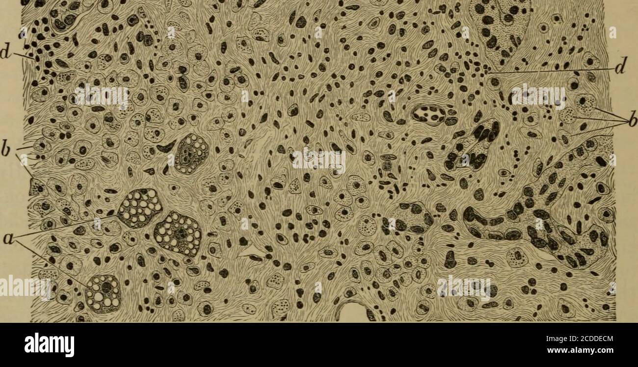
The elements of pathological histology with special reference to practical  methods . 0 « .V. Fig. 123.—Acute Yellow Atrophy of the Liver (from a spot  showing RedAtrophy), x 330. (Alum cochineal.)
The elements of pathological histology with special reference to practical methods . 0 « .V. Fig. 123.—Acute Yellow Atrophy of the Liver (from a spot showing RedAtrophy), x 330. (Alum cochineal.)

Histology Of Digestive Organs Lab

Digital Liver Histopathology Library

Bile duct – Normal Histology – NUS Pathweb :: NUS Pathweb